 Powered by Bioz


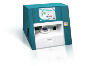
oCelloScope – Automated Microbial Live-Cell Imaging and Analysis
Automatically acquire and analyze images of your bacterial, fungus and yeast cells with real-time microscopic kinetic data
Description
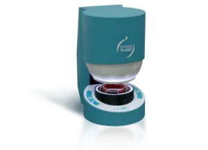
The oCelloScope is a powerful version of automated microscopy since it provides a live cell imaging solution. At its core, the oCelloScope can be used as a fungi microscope, an aspergillus microscope, a yeast microscope, and a fungal spore microscope. In other words, it is a live cell imaging microscope.
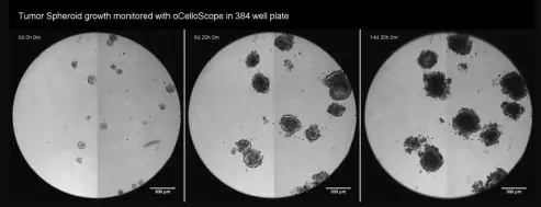
The ability of the oCelloScope to conduct microscope time lapse data collection and document it using video microscopy, makes it a powerful tool to evaluate bacterial growth kinetics as well as general microbial growth kinetics all in a multiwell format.
Do you know how your microbial cells develop?
The oCelloScope, as a version of automated microscopy, enables researchers to get more information and insight about anti-fungal treatments and antibiotic resistance. The oCelloScope, as a live cell imaging microscope, will automatically acquire and analyze images within standard microtiter plates, and will free up researchers’ time, thereby increasing sample throughput and delivering richer data.
The oCelloScope is a unique live cell imaging solution (for sensitive and detailed monitoring of biological growth and development. The oCelloScope system integrates 3 key features:
- Unique proprietary scanning technology, FluidScope™
- Powerful image analysis software
- Precision mechanical engineering
The 3 key features used together provide detailed information, enabling researchers to set up high-throughput testing and robust microscope time-lapse studies.
How can you benefit from live-cell image analysis with oCelloScope?
By combining a continuous real-time live cell imaging solution with powerful image analysis software, the oCelloScope facilitates investigations into natural preservatives, antimicrobial treatments, and antibiotic resistant organisms. Key capabilities of the oCelloScope include:
- Microscope time lapse – ensures that you will never miss a data point again
- Ability to capture and profile time dependent events
- Analyzes microbial growth and development
- Visualize and validate results with images and video microscopy
- Evaluates bacterial growth kinetics as well as general microbial growth kinetics
- Determines the minimum inhibitory concentration for antimicrobial treatments and natural preservatives
- Determines antifungal and antibacterial modes of action
Benefits
Speed
- 250 times more sensitive than a plate reader (OD)
- Measure and visualize down to 5 x 10³ CFU/ml
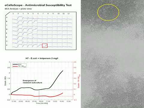
- Growth kinetic MIC results in few hours Vs 16-20h using BMD
- FluidScope™ technology scans a full 96-well plate in less than 3 minutes
Early Phase Morphology
- Compare growth kinetic curves with images from each time-point
- Discover microorganism antibiotic resistance adaptation strategies
- Microscope time lapse feature enables researchers to capture and quantify morphological changes over time
- Spheroplasts, Filamentation, Co-aggregation, Fungal spore germination
Value
- Full flexibility – use your standard microtiter plates
- You get full software package to use on multiple PCs
- No expensive annual service contract
- Competitive pricing. Rent or Purchase – just ask for a quotation
oCelloScope Workflow
With the oCelloScope functioning as a comprehensive instrument that incorporates the elements of a fungi microscope, an aspergillus microscope, a yeast microscope, and a fungal spore microscope, along with the powerful UniExplorer software, your research is supported day and night.
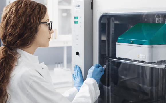
Make your experiment in any environment
- As a flexible, live cell imaging solution, the oCelloScope fits inside your incubator or anaerobic chamber
- Compatible with standard microtiter plates, from 6 to 96 wells
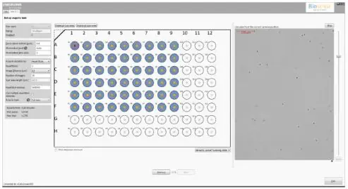
Setup automated acquisition and analysis
- Easy-to-use user interface for simple and flexible setup of your experiment
- Auto-focus and auto-illumination secures high quality data
Acquire images for as long as you like
- Microscope time lapse feature allows automatic acquisition of images for hours, days, or weeks using video microscopy.
- Use UniExplorer’s continue mode for running multiple long-term experiments simultaneously
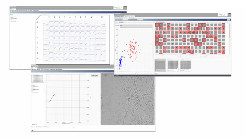
View and Analyze in real-time
- Follow you experiment in real-time with visualization of images and analysis
- Get an instant overview of your full 96-well plate
- Analyze single cells and mixed cultures
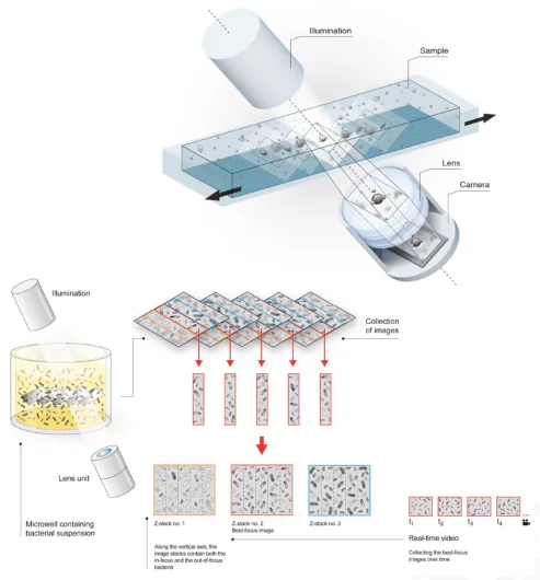
FluidScope™ scanning technology
As an element of automated microscopy, oCelloScope is based on the unique optical scanning technology FluidScope™ which combines optical techniques such as phase contrast, brightfield and confocal-like microscopy.
The OCelloScope, a live cell imaging microscope, which incorporates the advanced image processing algorithms of the FluidScope™ provides:
- Fast scanning of a volume in the sample
- Greater freedom of operation with multiple Z-layers acquired simultaneously
- Detailed 3D information at a single cell level
- Requires no pre-treatment, staining, or additional reagents
Measurement Specifications
| Sample objects | Most objects in (semi) transparent substances/liquid/agar can be analyzed, e.g.: Mammalian cellsBacteriaYeastFungiInorganic particles (crystals) |
| Sample matrix | Liquid and agar |
| Detectable density (sample concentration) | From 103 objects/mL |
| Detectable object size | 0.5 μm – 1 mm |
| Maximum scanning speed | 2 minutes 36 seconds (96-wells) |
| Optical magnification | 4x |
| Optical resolution | 1.3 μm |
| Camera | 5 Mpx, CCD, pixel size: 2.2 μm |
| Optical principle | FluidScope™ (patent pending) |
Environmental Specifications
| Operating temperature | 10 – 40ºC |
| Operating humidity | 20 – 93% RH |
Dimensions
| Dimensions (D x W x H) | 450 x 260 x 250 mm (with open lid: H = 550 mm) |
| Weight | 9.6 kg |
| Connectors | RJ45 (Ethernet) |
oCelloScope Microbiology Applications
oCelloScope Publications
Download Center
View Video